
Tissue Membrane Inspection
Inspecting every human tissue membrane allograft for defects, confirming its size, and recording the result before the graft is released.
The challenge
A regenerative-medicine manufacturer produces human tissue membrane allografts. Before a graft can be released, it has to be checked: that it is free of defects, and that it is the size it is supposed to be.
That inspection was being done by eye. A membrane graft is thin, translucent, and irregular, and the defects that disqualify one, a trapped bubble, a hole, a thin spot, a bit of foreign matter, are easy to miss and easy to judge differently from one inspector to the next. Doing it by eye is slow, it is inconsistent between people, and it leaves a thin paper trail behind a product that needs a strong one. On top of that the manufacturer makes many different graft products, each with its own size and its own idea of what counts as acceptable.
What we built
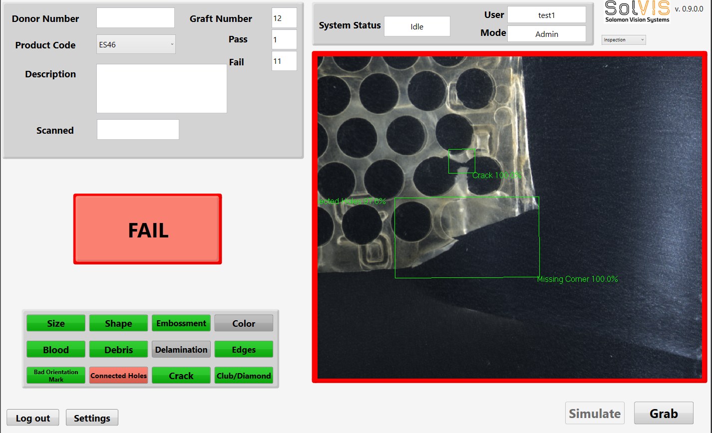
We developed the vision system that inspects the graft. Each membrane is imaged, and the software runs it through two stages. First, conventional machine-vision tools find and align to the graft in the image, so every inspection starts from the same reference regardless of how the piece was laid down. Then a deep-learning model, trained on real grafts, examines the tissue and flags defects, classifying what it found rather than just marking that something is there.
The graft is divided into a grid, and a defect is reported by type and by the grid cell it sits in, so a result is specific: not just "this one failed," but a bubble at a named location on the piece. The same pass measures the graft's width and length, so the dimensional check happens together with the defect check rather than as a separate step.
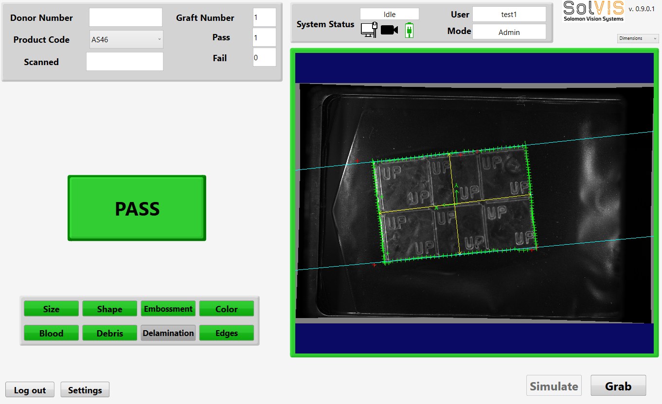
Because the manufacturer runs many products, the system is recipe-driven. Selecting the product loads the right expected size and the right trained inspection for that graft, so one system covers the whole product range instead of one build per SKU. The operator works from a single screen: choose the product, enter the piece's details, inspect, and the result comes back with any defect drawn on the image.
A record for every graft
For a medical product the inspection is only half the job; the evidence is the other half. Every inspection can be saved, both the image and the result, and the system aggregates the statistical data behind those decisions rather than discarding it. What was inspected, what was found, and where, is captured for each graft, which is exactly the kind of record a regulated tissue product needs and a manual visual check never produced.
Built to repeat
The manufacturer did not want one machine, they wanted several, running the same inspection to the same standard across more than one line. The system was designed so the software and its trained inspections carry over from the first build to the next, with retraining only where a new machine's imaging calls for it, so every unit inspects to the same bar rather than each being tuned by hand.
Capabilities used
Machine vision, deep-learning defect classification, dimensional measurement, operator GUI development, recipe-driven multi-product handling, image and statistical record-keeping, and repeatable deployment across multiple machines.
The system
Need an inspection that has to be right, and provable?
Engineer-led, customer-direct, backed by 24/7 support.
(423) 567-3755Talk to an engineer